Optical Imaging & Vital Microscopy Core
Baylor College of Medicine

We are a Light Microscopy Core Facility Specializing in Optical Sectioning Tools for Fluorescence Microscopy

The mission of the Optical Imaging & Vital Microscopy (OiVM) Core is to give our investigators the tools and technical expertise they need to obtain reliable, reproducible scientific data using state-of-the-art microscopy modalities.  We have over 20 years of experience educating, training and assisting scientists with experiments using the latest cutting edge imaging methodologies.

Our core specializes in vital and intravital imaging of cellular processes within cells, intact tissue explants, developing embryos and functioning organs within the live animal. Our users are focused on a variety of applications such as understanding cell migration, optimizing angiogenic therapies, how blood flow influences development and cancer, immune cell recruitment, stem cell-niche interactions and cancer metastasis.

We Provide Everything You Need to Streamline Your Experiments

Experimental Consultation

We provide guidance for all types of imaging experiments and we can help you select the instrument that will best suit your needs.

Training & Tutorials

We offer expert training and instruction programs tailored for beginners as well as experienced investigators.

Independent Use

Once our in-depth training is complete, the core is available 24/7 on a first come, first served basis

Available Technologies

The following microscopy methods are available through the Optical Imaging & Vital Microscopy Core. See information on each below:

Confocal Microscopy

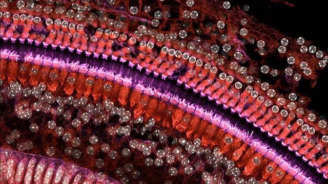
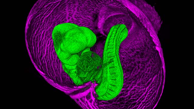
Learn more about how our confocal microscopes can improve your fluorescence images.

Lightsheet Microscopy

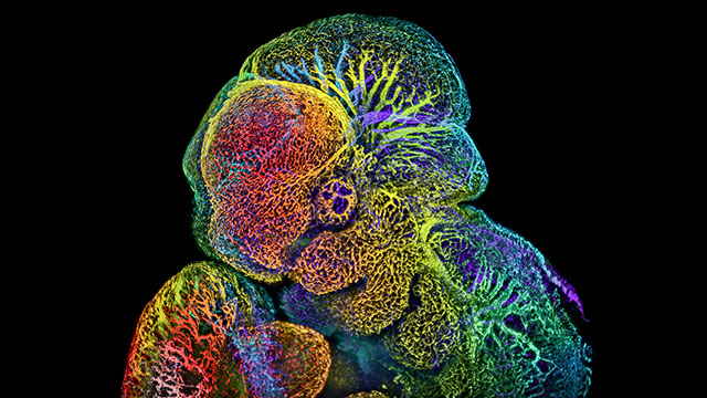
Learn more about how this novel 3D imaging technology can provide more insight into cleared whole mounts.

Multi-Photon Microscopy

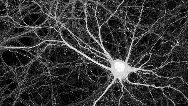
Learn more about how our 2-Photon microscopes can help you see deeper into intact tissue.

Widefield Microscopy

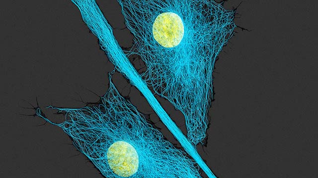
Learn more about how our widefield compound and stereo-Zoom microscopes can improve your fluorescence images.

μCT Imaging

Learn more about how μCT technology can generate 3D images from unlabeled samples.

Tissue Clearing

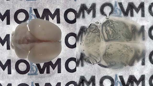
Learn more about how our X-CLARITY based tissue clearing services can render your in-tact organ systems transparent to light!

Supporting the Microscopy Needs of the Baylor College of Medicine and the Greater Texas Medical Center Community

Request Access to OiVM Core

Please fill out your contact information:
Please select either unassisted or assisted use:
Please check the boxes for all the services you are interested in:
Describe your experiment in detail: