Widefield Microscopy

Fluorescence is the foundation of our core. The bulk of instruments available for high resolution imaging at the OiVM are based on fluorescence. Fluorescence microscopy is a type of imaging where the emission signal from fluorophores such as molecules, quantum dots, nanoparticles, etc. is used to visualize cells and structures on a microscopic scale.

Fluorophores typically used in biology are excited by a specific wavelength of light. This excitation event causes the fluorophore molecules to move from a resting state (s0) to an excited state (s1). Once a fluorophore reaches an excited state (s1) it will begin to relax and enter into an intermediate vibrational state. After entering this vibrational state, it will spontaneously emit light within some defined, usually longer, wavelength range.

We can then use a microscope outfitted with filters that separate out excitation wavelength from emission wavelength. Cameras and other detectors can then be used to image the location of the fluorescence emission within the sample. Microscopes can be configured with many filter sets designed to image multiple fluorophores automatically. On our more advanced confocal systems, spectral array detectors are used to image up to 10 fluorophores simultaneously.

For a more comprehensive discussion of how fluorescence microscopy can benefit your research, please see our ‘Introduction to Fluorescence Microscopy’ video below.

For a list of the widefield microscopes available in the OiVM Core, please see our links below.

Video Coming Soon

Available Widefield Microscopes

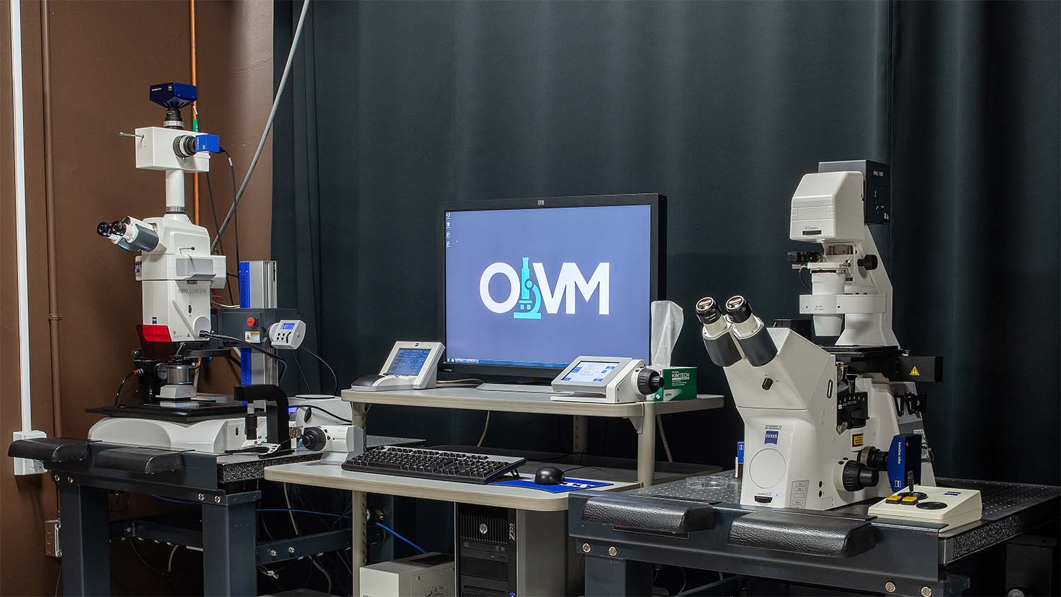
Carl Zeiss AxioObserver/AxioZoom Workstation

The AxioObserver/AxioZoom Workstation is a widefield platform that hosts both an inverted compound microscope AND an upright Zoom microscope.