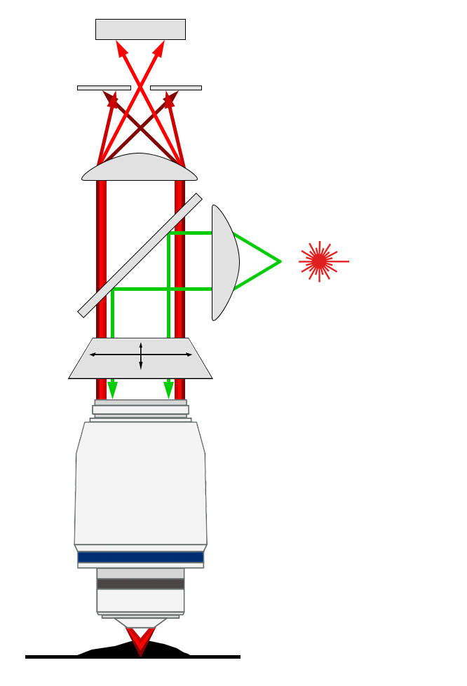
This technique leads to a significant improvement in axial resolution as well as contrast of the final image. Our confocal microscopes can be used to image a diverse range of biological specimens at sub-micron resolution. Most of our microscopes are equipped with incubation chambers that allow the regulation of temperature and CO² for live cell imaging over long periods of time.
For a more comprehensive discussion of how confocal microscopy can benefit your research, please see our ‘Introduction to Confocal Microscopy’ video below.
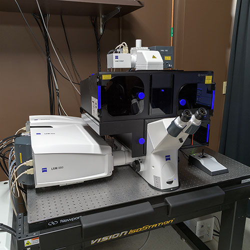
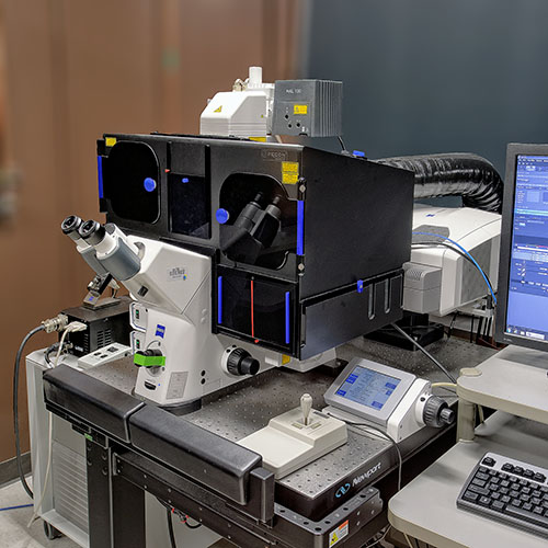
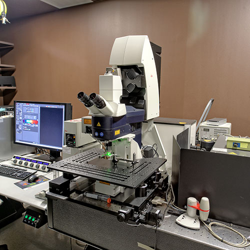
For a list of the confocal microscopes available in the OiVM Core, please see our links below.